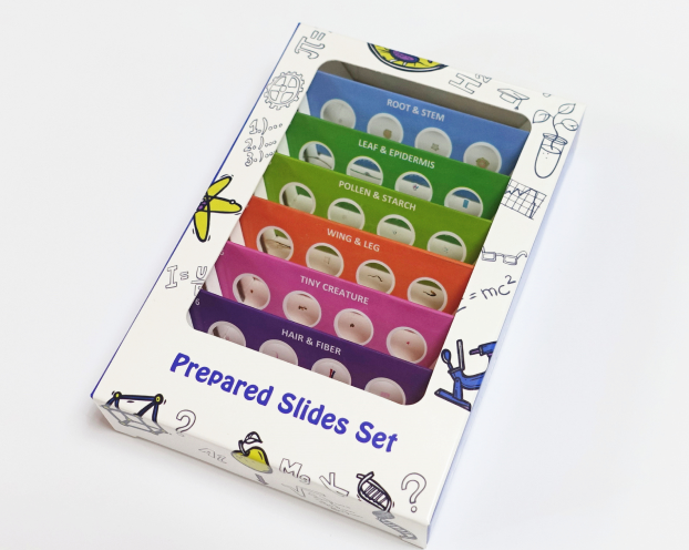
This page is not the official website. Generated by AI, does not constitute any advice. If needed, please click here to visit the corresponding official website.
Exclusively focused on researching and manufacturing high-quality prepared microscope slides since 1958. Trusted by educators, researchers, and institutions worldwide for consistency, reproducibility, and scientific accuracy.
Explore Products
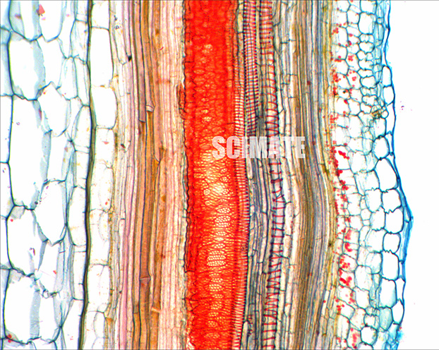
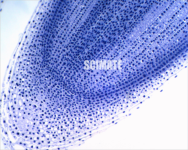
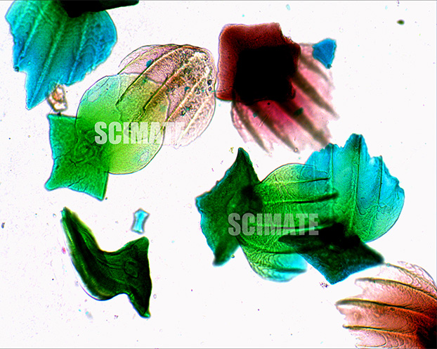
In the Scimate facility, manufacturing is seen as an art form. Using a unique staining technique, our skilled craftsmen bring their expertise and precision to every stage of production.
From tissue fixation to embedding, staining, sectioning, and mounting, each step is executed with the utmost care and precision. Scimate combines scientific knowledge with the artistry of craftsmanship to create high-quality prepared microscope slides that are consistent in quality, reproducibility, and reliability.
Each slide is meticulously crafted using advanced histological techniques and premium materials to ensure clarity, contrast, and educational value.
Wright-Giemsa stained peripheral blood smear showing erythrocytes, leukocytes, and platelets.
Longitudinal section showing active mitotic phases in plant meristematic tissue.
Erythrocytes with visible nuclei, ideal for comparative hematology studies.
Stratified squamous epithelium with dermal papillae and sweat glands.
Dicotyledonous leaf showing palisade mesophyll, spongy tissue, and vascular bundles.
Hematopoietic tissue showing developing blood cells at various stages.
Symbiotic nitrogen-fixing bacteria in plant root tissue.
Striated muscle fibers with visible sarcomeres and nuclei at periphery.
Respiratory cartilage showing chondrocytes in lacunae and matrix.
Secondary growth in gymnosperms with annual rings and resin canals.
Nephrons with glomeruli, proximal and distal convoluted tubules.
Chitinous structure with branching tracheoles and support veins.
Craftsmanship meets scientific precision in every slide we produce.
Each slide undergoes rigorous quality control to ensure anatomical correctness and staining fidelity for reliable educational and research use.
Our artisans combine decades of experience with precision techniques in sectioning, staining, and mounting for flawless results.
Sealed with durable mounting media to prevent fading and dehydration—slides remain clear and usable for decades.
We use a wide range of histological stains including Hematoxylin & Eosin (H&E), Wright-Giemsa, Toluidine Blue, Safranin, Fast Green, Methylene Blue, and specialized immunostains depending on the specimen and educational purpose.
Absolutely. Our slides are designed for both educational and research applications. They are durable, clearly labeled, and optimized for visibility under standard classroom microscopes.
All slides are permanently mounted using high-quality Canada balsam or synthetic resin to prevent drying and fading. Coverslips are sealed to ensure longevity and resistance to environmental factors.
Yes, we offer custom slide set development for schools, universities, and research institutions. Contact us with your curriculum or research requirements for a tailored solution.
Standard orders are processed within 3–5 business days. Custom sets may take 7–10 days depending on complexity. International shipping times vary by destination.
For inquiries, custom orders, or catalog requests, reach out to our team directly.
Notice for Buyers: If you are a buyer, the above information is generated by AI collection and does not constitute any advice. If necessary, please visit the corresponding official website.
For Domain Owners: If you are the domain owner and do not want to be included by fobcompany.info, please contact support@fobcompany.info via your corporate email to cancel. We will cancel your inclusion within 3 business days.
如果您是域名所有者 不想被fobcompany.info收录 请用企业邮箱联系support@fobcompany.info 取消收录 我们将在3个工作日取消您的收录
Service Request: If you are other domain and want to be included, please contact support@fobcompany.info
如果您是其他域名合作 也请联系support@fobcompany.info